Chiari malformation is a condition in which brain tissue extends into your spinal canal. It occurs when part of your skull is abnormally small or misshapen, pressing on your brain and forcing it downward.
Chiari malformation is uncommon, but increased use of imaging tests has led to more frequent diagnoses.
There are three types of Chiari malformation which depend on the anatomy of the brain tissue that is displaced into the spinal canal and whether developmental abnormalities of the brain or spine are also present.
Chiari malformation type I develops as the skull and brain are growing. As a result, signs and symptoms may not occur until late childhood or adulthood. The paediatric forms, Chiari malformation type II and type III, are present at birth (congenital). Being an adult surgeon I do not treat these two conditions. I shall discuss the Type 1 below.
Chiari Malformation Type 1
In Chiari malformation type I, signs and symptoms usually appear during late childhood or adulthood.
Headaches, often severe, are the classic symptom of Chiari malformation. They generally occur after sudden coughing, sneezing or straining. The other symptoms could include
- Neck pain
- Unsteady gait (problems with balance)
- Poor hand coordination (fine motor skills)
- Numbness and tingling of the hands and feet
- Dizziness
- Difficulty swallowing, sometimes accompanied by gagging, choking and vomiting
- Speech problems, such as hoarseness
Less often, people with Chiari malformation may experience:
- Ringing or buzzing in the ears (tinnitus)
- Weakness
- Slow heart rhythm
- Curvature of the spine (scoliosis) related to spinal cord impairment
- Abnormal breathing, such as central sleep apnoea, which is when a person stops breathing during sleep
As you can see a lot of the symptoms are non-specific and difficult to objectively assess.
Possible associated conditions
In some people, Chiari malformation can become a progressive disorder and lead to serious complications. In others, there may be no associated symptoms, and no intervention is necessary. The complications associated with this condition include:
- Hydrocephalus. An accumulation of excess fluid within your brain (hydrocephalus) may require placement of a flexible tube (shunt) to divert and drain the cerebrospinal fluid to another area of your body. The symptoms that are exacerbated by hydrocephalus is headache, nausea and vomiting.
- Syringomyelia. Some people with Chiari malformation also develop a condition called syringomyelia, in which a cavity or cyst (syrinx) forms within the spinal column.
- Tethered cord syndrome. In this condition, your spinal cord attaches to your spine and causes your spinal cord to stretch. This can cause serious nerve and muscle damage in your lower body. This stretch is often seen as the cause for the brain to herniate into the spinal canal.
Diagnosis
A review of your medical history and symptoms and a physical examination will be necessary. In addition, a few imaging tests may be required to make the diagnosis.
The tests will include
- Magnetic resonance imaging (MRI). An MRI is often used to diagnose Chiari malformation. This safe, painless test produces detailed 3D images of structural abnormalities in your brain that may be contributing to your symptoms. It can also provide images of your cerebellum and determine whether it extends into your spinal canal.
An MRI can be repeated over time, and it can be used to monitor the progression of your disorder.
- Computerized tomography (CT) scan. When finer details of the bony anatomy of the region is required a CT scan is particularly useful.
Treatment
Treatment for Chiari malformation depends on the severity and the characteristics of your condition.
If you have no symptoms, your doctor likely will recommend no treatment other than monitoring with regular examinations and MRIs.
When headaches or other types of pain are the primary symptom, your doctor may recommend pain medication.
Reducing pressure with surgery
Surgery is contemplated only when there is a symptomatic Chiari malformation. The goal is to stop the progression of changes in the anatomy of your brain and spinal canal, as well as ease or stabilize your symptoms.
When successful, surgery can reduce pressure on your cerebellum and spinal cord and restore the normal flow of spinal fluid.
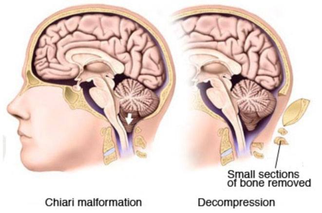
In the most common surgery for Chiari malformation, called posterior fossa decompression, or Foramen Magnum Decompression. your surgeon removes a small section of bone in the back of your skull, relieving pressure by giving your brain more room.
In many cases, the covering of your brain, called the dura mater, may be opened. Also, a patch may be sewn in place to enlarge the covering and provide more room for your brain. This patch may be an artificial material, or it could be tissue harvested from another part of your body such as your thigh.
Your doctor may also remove a small portion of the spinal column to relieve pressure on your spinal cord and allow more space for the spinal cord.
The surgical technique may vary, depending on whether a fluid-filled cavity (syrinx) is present, or if you have fluid in your brain (hydrocephalus). If you have a syrinx or hydrocephalus, you may need a tube (shunt) to drain the excess fluid.